3D Tomography reconstruction
Description
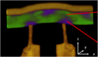
Two microscopy laboratories in Grenoble (CEA-Grenoble and Institut NÉEL) offer 3 3D tomography reconstruction techniques using transmission and scanning electron microscopy (SET = Scanning Electron Tomography) to characterize structure and/or chemistry in three dimensions at nanometric or atomic resolutions.
Volumes analyzed are typically of the order of 80 nm x 80 nm x 80 nm, and are extracted from bulk samples or larger nanodevices. Experimental measurements can be made from samples such as a deposition on a hole membrane or graphene membrane, but may also require the extraction of FIB thin films or tips.
Location
PFNC-CEA-Grenoble, Institut NEEL-Grenoble
Contacts
Platform managers: Jean-Luc ROUVIERE, Zineb SAGHI, Martien HERTOG
Associated targeted project coordinator: Pascale Bayle-Guillemaud
Technical characteristics
Chemical tomography (EELS-SET)
Uses electron energy loss (EELS) and the latest generation of direct-detection cameras to provide rapid 3D chemical analysis with near-nanometer resolution (CEA-Grenoble).
Contrast tomography (Nano-SET)
Uses a direct-detection camera and improved reconstruction algorithms (based on Machine Learning) to rapidly produce 3D structural maps with nanometer resolution (CEA-Grenoble).
Tomo-Ptychography (Ptycho-SET)
Uses a direct-detection camera and ptychography algorithms to reconstruct the device’s structure in 3D, at atomic scale (note that the thickness of the structure must be extracted below 40 nm) (Institut NÉEL and CEA-Grenoble).
3 steps are required for these three-dimensional characterizations:
- Sample preparation for TEM observation: FIB extraction or preparation of the structure to be analyzed,
- Electron microscope experiments,
- Data processing – SET-DIA’s essential contribution is the development of specific codes to accelerate reconstruction.
Associated targeted project